

Description
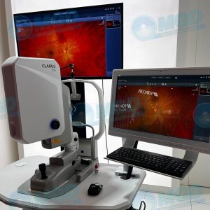
Nidek RS 330 Retina Scan-Duo
Nidek RS 330 Retina Scan-Duo incorporates new features enhancing screening and clinical efficiency in addition to user-friendly features that were incorporated from the previous model.
With the Retina Scan Duo 2, a 12 x 9 mm wide area OCT image can be acquired. The retina map captures both the macula and disc in a single shot. Normative database analysis is performed for both the disc and macula on a single OCT image, facilitating efficient diagnostic screening.
The Retina Scan Duo 2 incorporates a new image enhancement technique for denoising B-scan images. This function was released in December 2020 as B-scan Denoising Software for the NIDEK OCT series, which provides high-definition images comparable to a multiple-image-averaging technique, with deep learning of a large data set of images averaged from 120 images. By generating a high definition image from a single-frame image, this function decreases image acquisition time, ensuring greater patient comfort.
Combining image denoising with the retina map results in high-definition wide field area OCT images in a single shot with faster image acquisition at speeds of 70,000 A-scans/s for regular OCT sensitivity.
Large area scan
The wide area macula centered scan is the key image of the Nidek RS-330 Retina Scan Duo. This equipment provides a color-coded guide map to compare the patient’s macular thickness with the expected standard. Your practitioner will be better able to make diagnosis thanks to the comprehensive layout and comparison by the large scan.
FEATURES
User-Friendly Interfaces for Two Capture Modes
3-D Auto Tracking and Auto Shot
Operation with Joystick for Flexible Alignment
Space-saving Unit
HD Image Averaging (max. 50 images)
Selectable OCT Sensitivity – ultra-fine, fine, regular
Enhanced Image
Wide Area Scan (12 x 9 mm) / Wide Area Normative Database
Multiple OCT Scan Patterns
12-megapixel CCD Camera
Stereo and Panorama Photography
Fundus Autofluorescence (FAF)*1
En face OCT
NAVIS-EX
Anterior Segment Adapter*4


Reviews
There are no reviews yet.